
Enlarged jejunal lymph node in a red-necked wallaby
Case information
A 1.5-year-old, castrated, male red-necked wallaby presented for a one-week history of inappetance, depression, and diarrhea. The primary veterinarian had administered penicillin to no effect. The wallaby was housed in a fenced, outdoor enclosure with limited exposure to wild animals. The owners had an indoor-only cat; while the wallaby was raised indoors throughout the fall and winter, it did not have direct contact with the cat. The wallaby was up to date on rabies and tetanus vaccinations, had no exposure to toxins, no known ingestion of foreign material, and no changes in diet or husbandry.
On presentation, a firm, tubular structure was palpated in the dorsal abdomen. The rest of the physical examination was within normal limits. Abdominal radiographs revealed equivocally gas distended intestines. Abdominal ultrasound revealed fluid-filled intestines, normal gastrointestinal motility, a small amount of free abdominal fluid, and an enlarged jejunal lymph node, which corresponded to the mass detected on palpation. A hemogram revealed a leukocyte count of 4.4 x 10³ cells/μL (International Species Information System; mean ± SD; 5.9 ± 2.6 x 10³/μL), consisting of 2.5 x 10³ neutrophils/μL (2.0 ± 1.2 x 10³/μL), 0.1 x 10³ band neutrophils/uL (1.0 ± 2.0 x 10³/μL) and 1.8 x 10³ lymphocytes/μL (3.3 ± 1.8 x 10³/μL). There was moderate toxic change in neutrophils and a few reactive lymphocytes were seen. The platelet count was 111 x 103/μL (183 ± 110 x 103/μL). Pertinent chemistry results are shown in the table below:
| Test |
Result
|
ISIS
|
| Sodium (mEq/L) |
130
|
140 ± 5
|
| Potassium (mEq/L) |
2.2
|
4.9 ± 1.3
|
| Chloride (mEq/L) |
85
|
97±5
|
| Urea nitrogen (mg/dL) |
55
|
25 ± 6
|
| Creatinine (mg/dL) |
1.5
|
1.1 ± 0.3
|
| Albumin (g/dL) |
2.3
|
4.2 ± 0.6
|
| Globulin (g/dL) |
3.4
|
2.4 ± 0.9
|
| Glucose (mg/dL) |
218
|
96 ± 27
|
| ALT (U/L) |
491
|
48 ± 27
|
| AST (U/L) |
898
|
95 ± 92
|
| ALP (U/L) |
1048
|
1062 ± 891
|
| GGT (U/L) |
27
|
40 ± 24
|
| Total bilirubin (mg/dL) |
0.3
|
0.2 ± 0.2
|
| Creatine kinase (U/L) |
25039
|
1221 ± 902
|
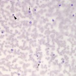
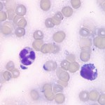
An aspirate was taken from the enlarged jejunal lymph node. View the photomicrographs then answer the questions below.
- What is your diagnosis based on the cytologic findings of the lymph node aspirate?
- How does this result explain the changes in the hemogram and chemistry profile?
- What additional tests are indicated?
|
|
|
Answer on next page