
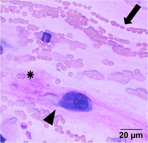
The red blood cells are lining up in rows within the mucinous material (arrow), with thicker strands of pink mucinous matrix material (*). Low numbers of individualized spindle cells were caught up within the matrix (arrowhead) (Wright’s stain, 50x objective).