
Facial mass on a goat
Case information
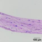
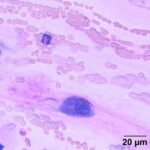
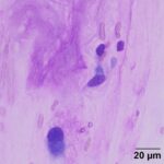
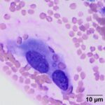
A 7-year-old male Nigerian Dwarf Goat presented with a mass on the left cheek. The owner reported the mass had been present for about two years, but had recently begun to increase in size. The lesion was described as a 2 cm round dermal mass. A fine needle aspirate of the mass was performed and smears were submitted to the Animal Health Diagnostic Center (AHDC) at Cornell University. The slides were stained in the laboratory and representative images are provided below.
Provided below are representative images of the fine needle aspirate.
|
|
|
|
|
Using the provided information, answer the following questions;
- What are your differential diagnoses for the pink background material in the images?
- What term is used to describe the arrangement of the red blood cells in this sample?
- What cellular features can be used to narrow down the differential diagnostic list?
Answers are provided on the next page.