
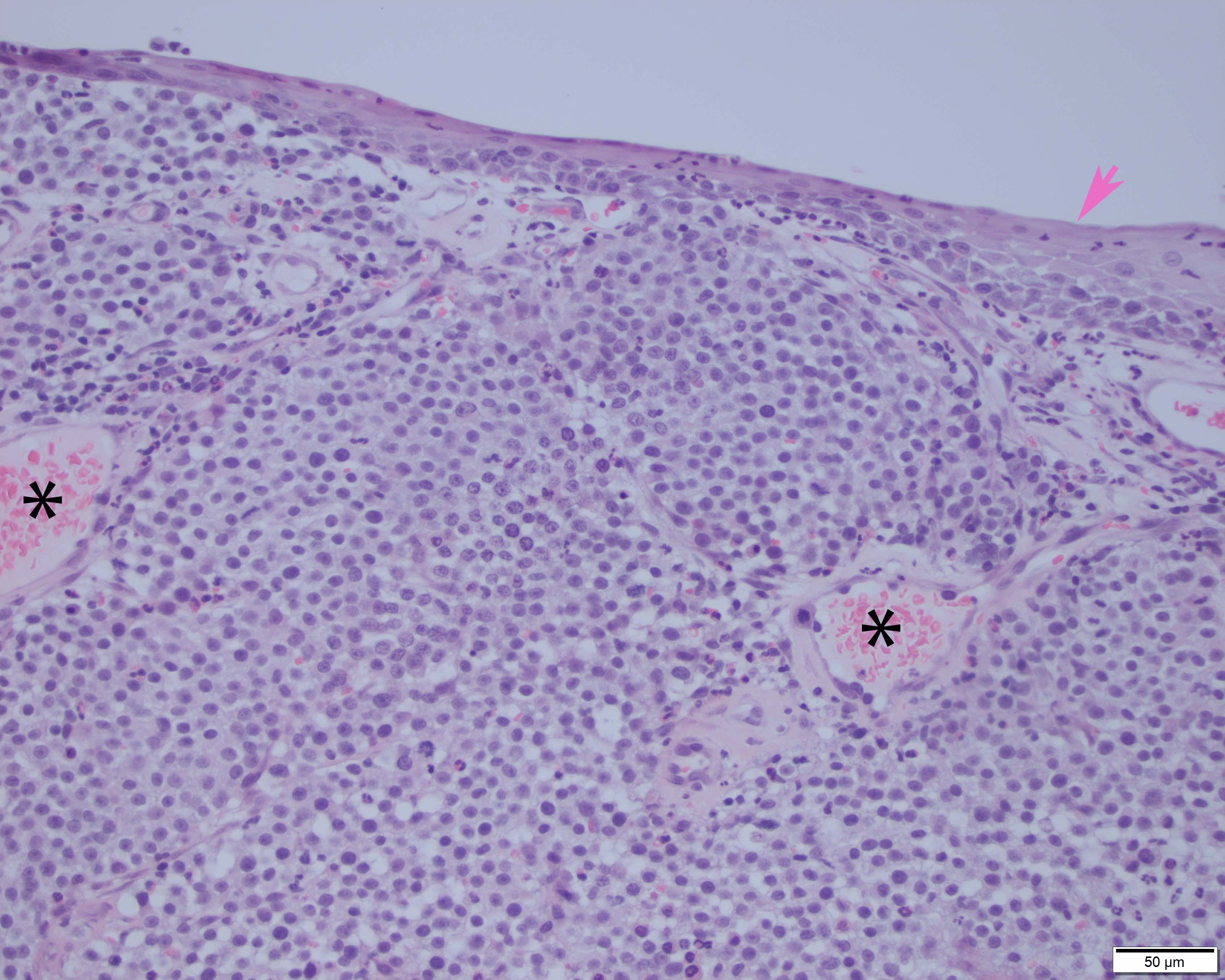
Figure 4: In the dermis underneath non-haired skin (pink arrow), there were large numbers of round cells in sheets, delineated by moderate amounts of fibrovascular stroma and peppered with blood vessels (asterisks). Focal areas of necrosis and hemorrhage (not shown) and heavy infiltrates of inflammatory cells were also present. H&E stain, 20x.