
Cauliflower-like mass at the base of the penis of a dog
Case Information
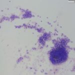
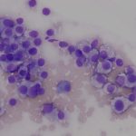
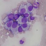
A 2-year-old castrated male mixed breed dog presented to the Cornell University Hospital for Animals (CUHA) Surgery Service for surgical revision of a previously amputated hind limb. The dog was rescued from the streets in Africa a few months prior and had partially amputated and infected right and left hindlimbs. The dog underwent a complete right hindlimb amputation with no complications. During post-operative care, a urinary catheter was placed and a 4.5cm x 3.4cm x 3.4cm cauliflower mass was observed at the base of the penis. The mass was friable, producing a mild serosanguinous discharge. Impression smears of the mass were taken and submitted to Clinical Pathology for microscopic examination.
Evaluate the provided representative images from the mass and answer the following questions:
- What is the top differential diagnosis?
- What is the significance of the other cells present?
- What is the main route of “infection”?
|
|
|
|
Answers on next page