
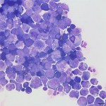
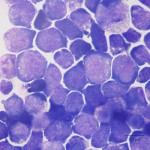
Photomicrographs of cerebrospinal fluid from a dog
Case information
An 8 year old intact male Labrador retriever presented for acute hind limb ataxia. The dog had a recent history of shifting limb lameness and left-sided facial paralysis that had partially resolved. The day prior to presentation, the dog had become lethargic and inappetent, and had an acute episode of pain that resulted in severe ataxia and hind limb weakness. At presentation, the dog was non-ambulatory and paraparetic. Proprioceptive deficits and decreased withdrawal response were noted in the hind limbs. Patellar reflexes were intact. The dog appeared painful on palpation of the thoracolumbar region of the spine. Evidence of cranial nerve deficits included the previously noted left-sided facial droop, positional ventral strabismus and absence of palpebral or menace responses of the left eye. The neurologic examination was consistent with multifocal disease involving the T3-L3 of the spinal cord and cranial nerves V, VII, and VIII. The remainder of the dog’s physical examination was unremarkable. Magnetic resonance imaging of the spine revealed hyperintensity in the epaxial muscles and hypointensities within several nerve roots. An infiltrative disease process was suspected to be occurring within the parenchyma of the spinal cord (T13-L3) and differential diagnoses included neoplasia, inflammation, and infection. Cerebrospinal fluid (CSF) collected from the lumbosacral space was colorless and slightly cloudy. The nucleated cell count was 4277 cells/uL and total protein was 2340 mg/dL. Evaluate the representative photomicrographs and consider the following questions:s:
- What cell types can be found in normal CSF?
- What cell type comprises the majority of the cells in this dog’s CSF?
|
|
|
Answer on next page