
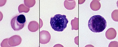
This image shows two types of nRBC and a lymphocyte from a dog. Left panel: A metarubricyte with red cytoplasm. This is the most typical form seen in animals. Middle panel: A polychromatophilic rubricyte with a very clumped nuclear chromatin, and purple cytoplasm (due to a combination of RNA and hemoglobin) with raggedy or ruffled outlines. Right panel: A lymphocyte with smoother chromatin and small amounts of slightly grainy blue cytoplasm with smooth borders (Wright’s stain).