
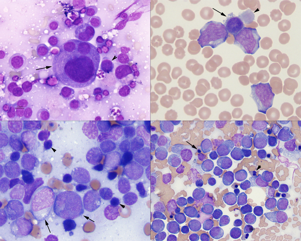
Upper right panel: Abnormal (dysplastic) megakaryocytes (arrow) and smaller cells with fine chromatin and high nuclear to cytoplasmic ratios that are presumptive megakaryoblasts (arrowhead) in a lymph node aspirate from a dog with acute megakaryoblastic leukemia (Wright’s stain, 50x objective). Upper right panel: Three myeloblasts (presumptive megakaryoblasts), a micromegakaryocyte (arrow) and dysplastic large platelet (arrowhead) in the blood of a dog with acute megakaryoblastic leukemia (different dog from that shown in the upper left panel) (Wright’s stain, 100x objective). The ruffled edges to the micromegakaryocyte, with the light gray-purple cytoplasm (resembling platelet cytoplasm) support a megakaryocytic lineage for the abnormal cell. Lower left panel: Myeloid blasts (arrows) infiltrating a peripheral lymph node in a dog with acute myelomonocytic leukemia. The cells are seen in a background of residual small lymphocytes (arrowheads), indicating an infiltrative population versus a primary lymphoid tumor. Some blasts have pleomorphic nuclei or increased amounts of light blue cytoplasm and they do vary in size, features suggesting (but not definitive for) myeloid lineage (Wright’s stain, 100x objective). It would be easy to mistake this for lymphoma. Lower right panel: Proerythroblasts (rubriblasts) dominate in a bone marrow aspirate from a cat with acute erythroid leukemia. Erythroid blasts have deep blue slightly grainy cytoplasm and uniformly round nuclei. Note the nucleated red blood cells (arrow), some of which are dysplastic (arrowhead) (Wright’s stain, 50x objective).