
Canine Distemper
Large aggregates of viral nucleocapsid particles can form in erythrocytes, leukocytes, and epithelial cells in many tissues during the acute phase of infection with Canine distemper virus, resulting in cytoplasmic inclusions visible by light microscopy. Their presence in blood is transient; they are very rarely encountered but are pathognomonic when identified.
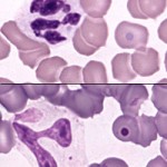
Shown at the right are two smears from a dog with distemper, the upper one stained with Wright’s stain and the lower with Hemacolor® or Diff-quik. The viral inclusions, present in each smear in both a red cell and a leukocyte, stain much more darkly with Hemacolor and other quick hematology stains; this facilitates their detection.