
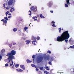
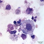
Photomicrographs of a urine sample from a dog
Case information
A 6 year old castrated male Akita presented with a one year history of persistent polyuria and polydipsia, that was unresponsive to antibiotic (Amoxicillin) treatment. The dog was difficult to examine, but appeared painful on abdominal palpation. Ultrasonographic imaging showed a diffusely thickened bladder wall. A urine sample was collected by cystocentesis and submitted for urinalysis and cytologic evaluation. Urinalysis results and representative cytologic images (from a cytospin smear of a urine sediment) are provided below.
| Urinalysis results | ||
| Test | Result | Units |
| Volume | 2 | mL |
| Color | Medium yellow | |
| Turbidity | Slightly cloudy | |
| Specific gravity | 1.016 | |
| pH | 8.0 | units |
| Protein | 15 (trace) | mg/dL |
| Heme | 1+ | |
| RBC | 5-20 | /HPF |
| WBC | <5 | /HPF |
| Bacteria | None seen | |
| Epithelial cells | Moderate | |
| Sperm | None seen | |
Evaluate the representative photomicrographs below and consider the following questions:
- Interpret the results of the urinalysis.
- Identify the labeled cells in Figure 1.
- Is a causative agent evident?
- what other tests are indicated on the urine?
|
|
|
Answer on next page