
Cutaneous mass from a cat
Case information
A 12 year-old female spayed cat presented for evaluation of a mass on the dorsal right cervical area. The mass was first noticed by the owners 3 weeks after a vaccine against Rabies was applied on the same location.
On presentation, the cat was bright, alert, and responsive and all her vital parameters were within normal limits. On physical examination, a 2.6 x 2.0 x 1.3 cm subcutaneous and movable mass on the right aspect of the dorsal cervical region was palpated. The rest of her physical examination was unremarkable.
Blood samples were obtained and submitted for a complete blood cell count and biochemistry panel and only revealed a mild hyperproteinemia (8.3 g/dL, reference interval, 5.9-7.5 g/dL). Thoracic radiographs and abdominal ultrasound were unremarkable. A computed tomography (CT) revealed a small non-discrete lesion above her right cervical area. A fine needle aspirate of the mass was obtained and submitted for cytologic evaluation.
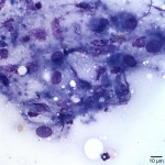
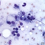
Examine the representative images that are provided below and answer the following questions.
- How would you classify the inflammation present?
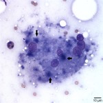
- What is the most likely origin of the pale to medium purple material indicated by the arrows in Figure 3 (also present in Figure 1 and Figure 2)?
- Given the clinical history, signalment and cytologic findings, what is the most likely cause of the inflammation?
|
|
|
|
Answers on next page