
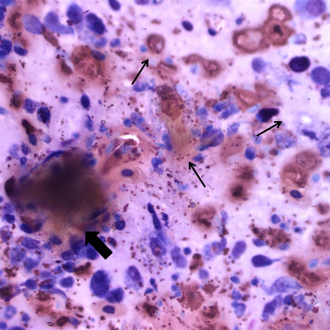
Figure 1B. Large numbers of heterophils and macrophages are present, with copious urate crystals admixed (thin arrows). A dense aggregate of urate crystals takes on a more brown hue than the individual crystals (thick arrow). Wright’s stain, 500x.
Figure 1B. Large numbers of heterophils and macrophages are present, with copious urate crystals admixed (thin arrows). A dense aggregate of urate crystals takes on a more brown hue than the individual crystals (thick arrow). Wright’s stain, 500x.
eClinpath helped 1.2 million visitors last year from 220 countries find important information on animal health. If you enjoy the site, please support our mission and consider a small gift to help us keep pace with its rapid growth. You can donate securely via PayPal or credit card. Thank you!
![]()