
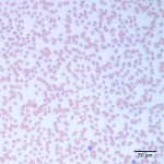
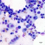
Photomicrographs of a blood smear and a bone marrow aspirate from a dog with ocular lesions
Case information
A 7 year old castrated male, Alaskan Malamute presented due to weight loss, lethargy, inappetance, intermittent epistaxis and ocular inflammation which began about two months prior. The patient initially presented due to clear discharge from his right eye and change of color of the eye (red, brown and gray discoloration). Treatment was initiated with oral and topical antibiotics but the eye lesions progressed and the patient became lethargic, inappetant and began exhibiting epistaxis. On his most recent presentation, his right eye was hyperemic with diffuse corneal cloudiness. A complete ophthalmic exam revealed chronic anterior uveitis and an indolent ulcer in the right eye and retinal detachments in the left eye.
Blood was drawn for a CBC and chemistry panel (Table 1) and an abdominal ultrasound was performed, which revealed a mildly enlarged liver and spleen. The patient was anesthetized for aspirates of his liver, spleen and bone marrow. A buccal mucosal bleeding time (BMBT) was done prior to these procedures to assess for bleeding potential given the history of epistaxis. The BMBT was prolonged at greater than 17 minutes (normal is less than 5 minutes). Based on this result, aspirates of the enlarged liver and spleen were canceled and only a bone marrow aspiration was performed. Evaluate the following abbreviated CBC and chemistry results (Table 1) and blood and bone marrow images (Figures 1 and 2, taken at 20X and 50X magnification) to answer the following questions:
| Table 1: Abbreviated CBC and chemistry results | |||
| Test | Result | Units | Ref Interval |
| Hct | 27 L | % | 41-60 |
| WBC | 3.3 L | thous/μl | 5.7-14.2 |
| Seg Neut | 1.5 L | thous/μl | 2.7-9.4 |
| PLT | 212 | thous/μl | 0.9-4.7 |
| Total protein | 9.2 H | g/dL | 5.9-7 |
| Albumin | 2.2 L | g/dL | 3.1-4.2 |
| Globulin | 7.0 H | g/dL | 1.9-3.6 |
| Ionized calcium | 1.45 H | mmol/L | 1.16-1.4 |
- What is the likely cause of the rouleaux formation on the blood smear? (Hint: You can confirm this by looking at the chemistry panel.)
- What is the predominant cell type in the bone marrow image below?
- What other test could be done to facilitate a definitive diagnosis in this case?
- Does this diagnosis explain the patient’s ocular signs?
|
|
|
Answer on next page