
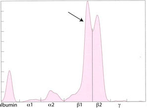
Electrophoretogram from a dog with an IgA-secreting multiple myeloma. Two monoclonal peaks are seen (arrow). Very little protein is observed in the γ-region suggesting concurrent immunodeficiency of the other immunoglobulin classes. The albumin peak is also low, but this is only in proportion to the large increase in the IgA. The dog actually had a normal albumin concentration when the % value (area under the curve) was converted to an absolute albumin concentration (g/dL) (by multiplying by the total protein concentration from the chemistry analyzer) and compared to our reference interval for albumin (this was because the total protein was quite high in this dog, i.e. 15 g/dL).