
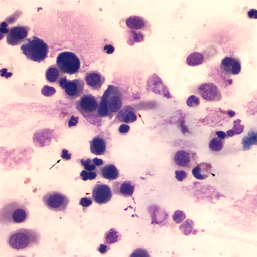
Note the respiratory epithelial cells, which range in appearance from cuboidal to columnar (red arrow). The cells have long wispy “tails” at one end and can mimic spindle cells but the opposite end is usually squared off and blunted. There are increased inflammatory cells, consisting of non-degenerate neutrophils (black arrow) with macrophages (red arrowhead) and eosinophils (black arrowhead) (modified Wright’s stain, 50x objective).