
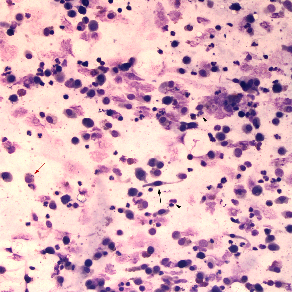
The tracheal wash sediment smears contained small amounts of mucus with respiratory columnar epithelial cells (black arrow) and increased inflammatory cells, consisting of macrophages (red arrow), some of which were binucleated, neutrophils (black arrowheads) and fewer eosinophils (modified Wright’s stain, 20x objective).