
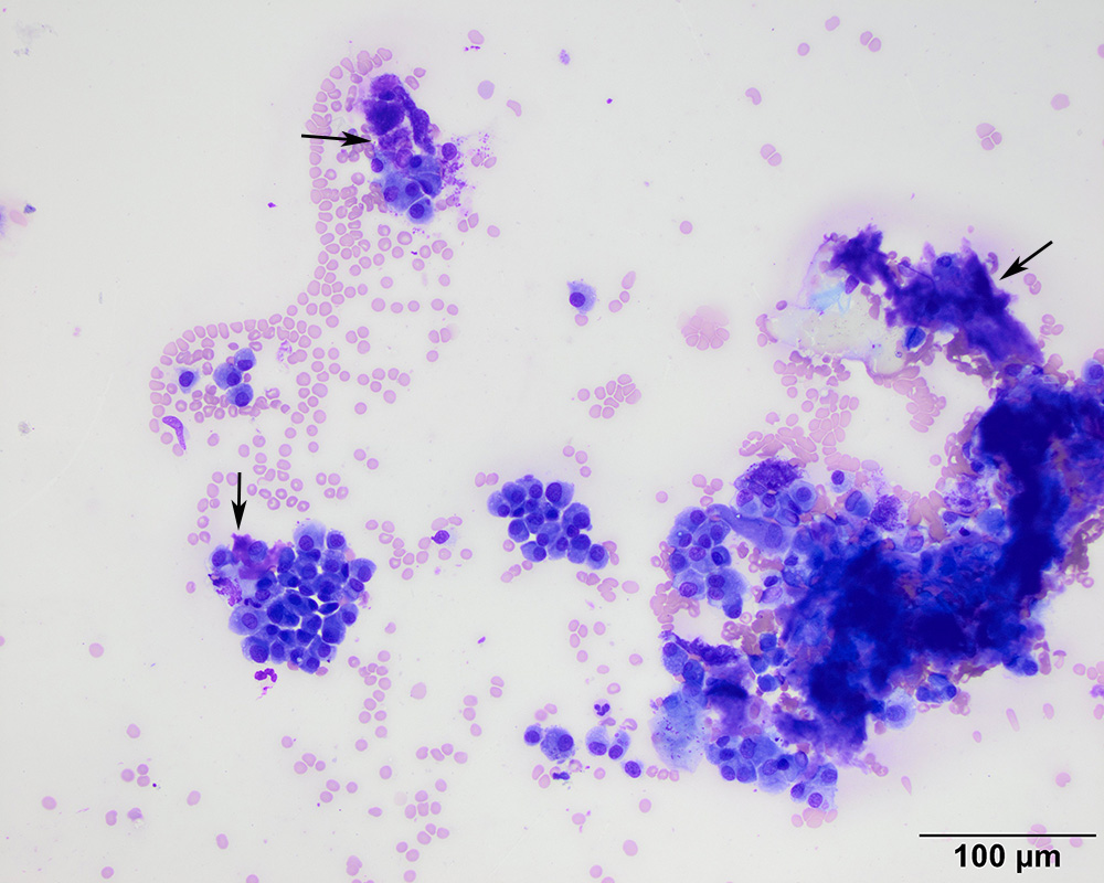
The aspirate consisted of numerous aggregates and individualized round cells with cytologic features of plasma cells. The cells were associated with a chunky to smooth purple extracellular matrix, compatible with amyloid (arrows) (Wright’s stain, 20x objective).