
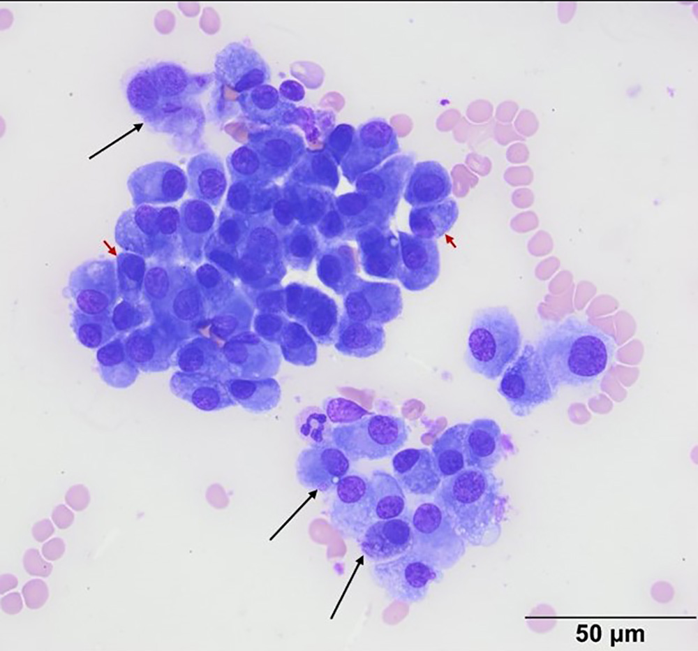
Non-cohesive aggregates and individualized plasma cells with cytoplasmic granulation of variable sizes (long black arrows) comprise the tissue cells in the aspirate. Several of the cells have distinct perinuclear clear zones and dark blue cytoplasm with eccentric nuclei and clumped chromatin (short red arrows), features characteristic of well-differentiated plasma cells (Wright’s stain, 50x objective).