
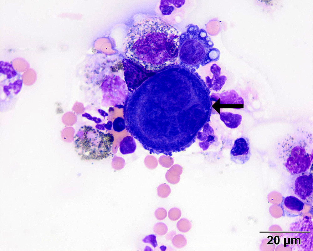
This image showcases a large reactive mesothelial cell, with deep blue cytoplasm and multiple nuclei (arrow), intermixed with malignant melanocytes, melanophages, neutrophils, and blood. (Wright’s stain, 100x objective).
This image showcases a large reactive mesothelial cell, with deep blue cytoplasm and multiple nuclei (arrow), intermixed with malignant melanocytes, melanophages, neutrophils, and blood. (Wright’s stain, 100x objective).
eClinpath helped 1.2 million visitors last year from 220 countries find important information on animal health. If you enjoy the site, please support our mission and consider a small gift to help us keep pace with its rapid growth. You can donate securely via PayPal or credit card. Thank you!
![]()