
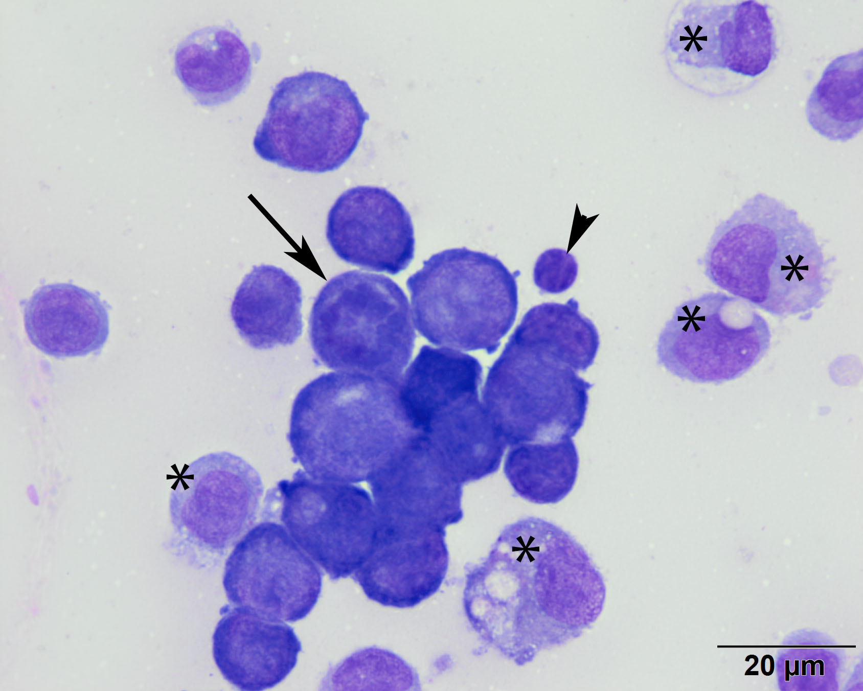
High power image showing morphologic features of neoplastic cells present in the center of the image. Rare mitotic figures were seen (arrow). Other cells present include mostly macrophages (*) and rare small lymphocytes (arrowhead). (Wright’s stain, 100x objective)