
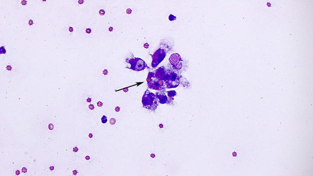
A group of macrophages, demonstrating cytophagic activity, including an erythrophage (arrow). Unlike the tumor cells, the macrophages contained less of the discrete margined vacuoles and more “phagocytic” vacuoles, which contain material within them and have less distinct margins (modified Wright’s stain, 50x objective, sediment smear).