
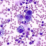
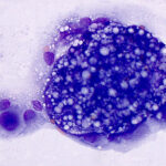
Pleural fluid from a 2 year old dog
Case Information
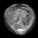
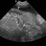
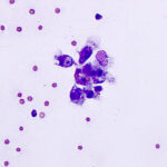
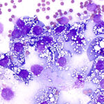
A 2 year old intact female mixed breed dog presented for evaluation of a bicavitory (pleural and peritoneal) effusion after showing mildly reduced activity. The dog was clinically healthy up until this time but had suffered from abnormal heats (increased frequency, prolonged, split heats). Routine hemogram, biochemical and coagulation testing did not reveal any abnormalities. On abdominal ultrasonographic examination, the uterus was enlarged and dilated and a 5.8 x 4.6 cm mass was noted next to the right cranial uterine horn (Figure 1). The left ovary was normal in appearance. Computerized tomography of the abdomen and thorax revealed a large amount of peritoneal and pleural fluid. A large cavitated nodular mass (10 x 11 x 14 cm, Figure 2), contiguous with the dilated right uterine horn, was seen caudal to the right kidney and mesenteric lymph nodes were enlarged. There was a concurrent nodular spiral pattern in the omentum. In the thorax, a large mass (6 x 9 x 9 cm) was noted in the left mediastinum, along with a focal alveolar infiltrate in the right cranial lung lobe and enlarged sternal lymph nodes. The pleural fluid was aspirated and mailed into the Animal Health Diagnostic Center for cytologic examination (a direct smear was also provided with the fluid). The fluid was medium red and opaque, with a total nucleated cell count of 10,300/μL, red blood cell count of 234,000/μL and a total protein by refractometer of 2.7 g/dL. Sediment smears were prepared from the fluid and stained with modified Wright’s stain.
Examine the provided images from the stained sediment smears of the fluid, then answer the following questions:
- What cells can you identify in the smears?
- What is your cytologic diagnosis?
- How do you relate these findings to the identified abdominal mass?
|
|
|
|
|
Answers on the next page