
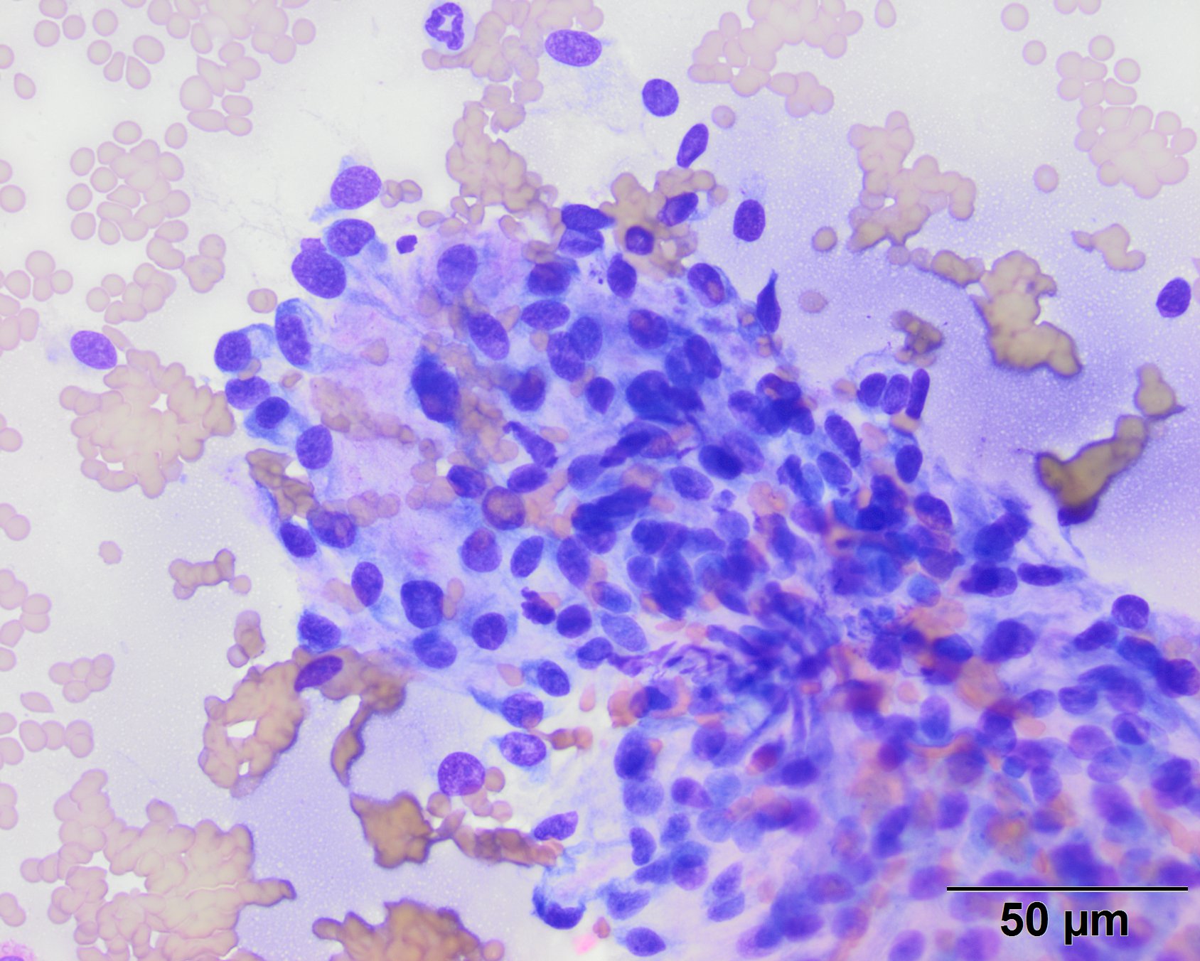
The spindle-shaped cells are embedded within light pink to purple extracellular matrix material and show mild anikaryosis and anisocytosis in this image (Wright’s stain, 50x objective)
The spindle-shaped cells are embedded within light pink to purple extracellular matrix material and show mild anikaryosis and anisocytosis in this image (Wright’s stain, 50x objective)
eClinpath helped 1.2 million visitors last year from 220 countries find important information on animal health. If you enjoy the site, please support our mission and consider a small gift to help us keep pace with its rapid growth. You can donate securely via PayPal or credit card. Thank you!
![]()